Research imaging in Edinburgh
Research by Edinburgh’s world-leading stroke experts, Professor Joanna Wardlaw and Professor Peter Sandercock, is helping more patients benefit from life-saving treatment.
Stroke is the second leading cause of death worldwide and a major cause of disability. It occurs when the supply of blood to the brain is interrupted or reduced due to blood vessel blockage (ischaemic stroke) or rupture (haemorrhagic stroke).
Stroke can be fatal and those who survive can experience a wide range of disabilities depending on which part of the brain has been affected and how quickly they are treated. Fast diagnosis and treatment not only saves lives, it can drastically improve recovery.
“ We’ve come a long way since the 1980s when there were no treatments for people who had just had a stroke and care was solely focussed on rehabilitation,” says Peter Sandercock, Emeritus Professor of Medical Neurology at the University.
Read more on this research and International Stroke Trials (IST)
Today, thanks to increased awareness of the early warning signs of stroke, victims are more likely to seek help straight away and to be admitted to specialist stroke units where they can be quickly scanned and receive drugs that reduce their chance of dying or becoming disabled.
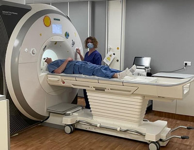
Imaging technologies, CT and MRI scanning, are the only way to determine what type of stroke a patient has had and therefore the treatment they will require. Joanna has been examining the most effective ways to use these technologies for assessing stroke patients and their eligibility for thrombolytic treatment.
Her team has shown that the most cost-effective way to manage patients with stroke is to use CT scanning straight away. Compared with MRI, CT scanning is faster, cheaper and easier to use in acute stroke. Although MRI scans can be very sensitive to the features of stroke, they may not always capture them.
“ For every 100 people who are clinically diagnosed with a mild stroke, 30 may not show the signs of it on an MRI scan, but they should still be treated,” Joanna says. “ It is important to recognise that clinical assessment forms the basis of stroke diagnosis; imaging should be considered a supporting tool.”
MRI scans can also be useful to detect signs of acute brain ischaemia in about a third of TIAs, or to determine the cause of stroke in patients who cannot be scanned until more than five days after stroke, when CT scans can no longer distinguish whether the patient has suffered an ischaemic or a haemorrhagic stroke. However, Joanna notes that there are many other highly justified uses of MRI such as cancer screening that are competing with its use in stroke.
Stroke research has largely focused on blood supply to the brain, particularly via large arteries leading from the heart, the muscular intracranial large arteries, large cerebral veins and venous sinuses, and more recently perforating arterioles, capillaries, and venules.
However, these blood vessels only serve part of the brain’s fluid management, nutrient delivery, and waste clearance system.
The other, and until recently largely neglected, aspect of brain fluid and waste management is the system that flushes the brain, draining interstitial and cerebrospinal fluid (CSF) and waste from the cranial cavity.
Details of this non–blood-vessel brain circulation are incomplete, but there is now enough clinical relevance for it to be the focus of this Advances in Stroke: Diagnosis and Imaging, particularly as key elements are now visible on routine magnetic resonance imaging (MRI).
Recently STV Scotland visited the University’s Edinburgh Imaging Facilities to find out about the dementia research taking place in Edinburgh for a dementia special, presented by Kaye Nicolson and aired on 21st April.
During their visit they interviewed Professor Joanna Wardlaw, Professor of Applied Neuroimaging, to discuss a new study involving magnetic resonance (MR) and retinal imaging, funded by the Alan Turing Institute and British Heart Foundation.
Professor Wardlaw discussed how, by scanning the brain, you are not able to see blood vessels. Therefore, researchers hope that studying blood vessels at the back of the eye, will give insight into what is actually happening to blood vessels in the brain.
The special also interviewed a study participant, whose blood vessels at the back of the eye were studied with a retinal scan and an MR scan at the Edinburgh Imaging Facility on Edinburgh BioQuarter.
The Edinburgh Imaging Facility is attached to the Royal Infirmary of Edinburgh building close to the Division of Clinical Neurosciences (DCN) on Edinburgh BioQuarter and delivers clinical & academic research imaging in humans.